A research team at Stanford University has developed a new deep imaging technology that can brightly illuminate buried tumors in the body of cancer patients. Using this technology, it is possible to observe the tumor progression in the non-invasive body more clearly than before.
According to a study published in the journal Nature Biotechnology on September 30, a new deep imaging technology useful for identifying cancer patient responses to immunotherapy and monitoring progress after treatment was released. The technology was developed by a research team led by Hongjie Dai, an applied physicist at Stanford University. He says that the technology relies on nanoparticles containing erbium, and erbium is one of the so-called rare earth elements as an element that is highly valued for its intrinsic property of emitting infrared light.
To cover the nanoparticles containing erbium with a chemically designed coating, the nanoparticles help dissolve in the bloodstream, reduce toxicity and promote faster excretion from the body. It is also explained that the nanoparticle coating acts like a guided missile against cancer cells, thanks to its usefulness in finding and attaching specific proteins in cells.
In the study, the mice ingested nanoparticles containing erbium were illuminated with low-power LED lights to explode blood vessels in the mice and successfully observed tumors or individual cells as target tissues with much higher resolution than conventional imaging techniques. The research team revealed that the developed deep imaging technology shows a level that cannot be achieved with conventional methods due to the combination of specificity, multiplicity, and spatiotemporal resolution.
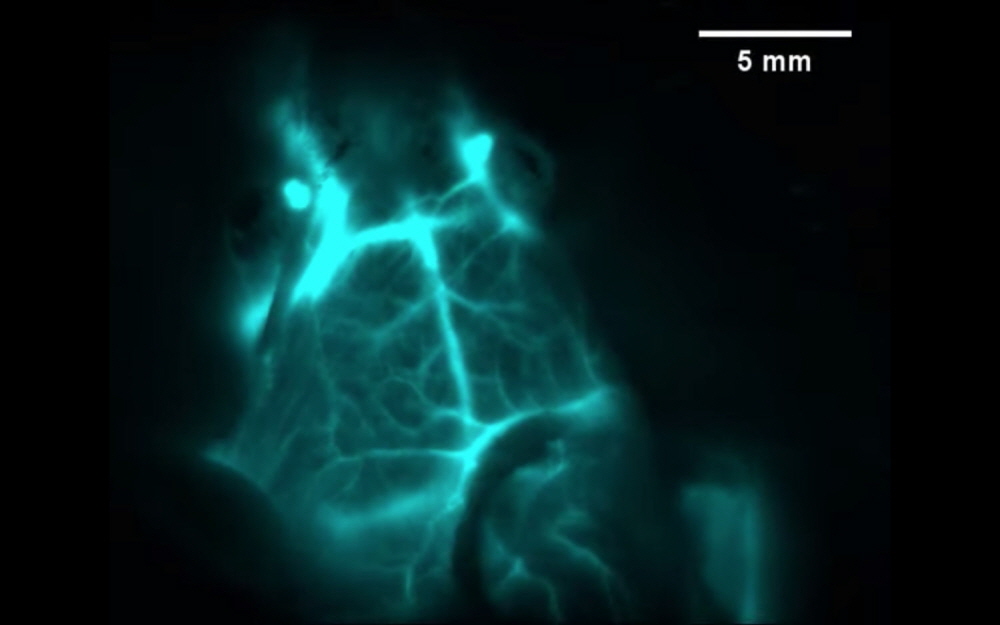
If you look at the image, you can see that the blood vessels of the brain glow in infrared rays by ingesting nanoparticles into a live mouse. Rat brain blood vessels glow in cyan. Through this approach, we can see the rat’s brain, and conventional methods only see the scalp.
Using this deep imaging technique, the research team succeeded in identifying tumors in mice susceptible to anticancer drugs that activate the immune system. Therefore, it is possible to provide a non-invasive method capable of specifying a site that responds to a drug without a test to collect a part of the lesion required by the existing method. In addition, erbium nanoparticles can be used to monitor the patient’s progress after cancer treatment and continuously monitor whether the tumor is responding to the drug or if the tumor is shrinking. The team explains that deep imaging techniques can help surgeons resect tumors more accurately or help biologists study the basic processes of cells. Related information can be found here .


Add comment